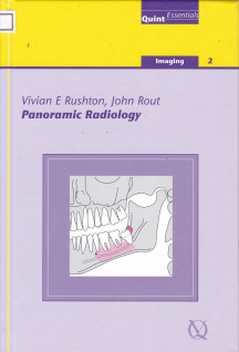
Panoramic Radiology
Rout, John (Author)
London
9781850970804
Quintessentials of dental practice
Not available
Not available
Rushton (University of Manchester) and Rout (Birmingham Dental Hospital) review the development of dental panoramic radiography during the past century, the anatomical structures that may appear on a DPR, and the principles of a quality assurance program for producing high quality diagnostic images with the minimum patient radiation dose. Example black and white radiographs of common conditions are provided.
148 pages
21 cm
Illustration, Index
Not available
| Access no. | Call number | Location | Status |
|---|---|---|---|
| 00016/26 | 617.607572 Rus P | Fakultas Kedokteran Gigi (FKG) | Available |